Jump To: What Is Ultrasound | Ultrasound in Medical | Manufacturing Ultrasonic | The Doppler Effect | Common Uses of Ultrasound | Kidney Stones | Malignant Tissue | Harmonic Scalpel
A transducer in medical settings converts electrical signals into ultrasound waves, or conversely converts ultrasound waves back into electrical signals. In medical ultrasound, piezoelectric transducers use lead zirconate titanate (PZT) ceramics as the active element that vibrates at high frequencies to produce ultrasound waves at scales of 10 MHz or higher for diagnostic and therapeutic procedures.
Transducers in medical practice enable ultrasonic imaging for visualizing internal organs, arteries, and tissues with high-resolution images of about 0.2 millimeters. Transducers in medical applications use the Doppler Effect to measure blood flow velocity within arteries, providing physicians with detailed diagnostic information for conditions affecting the abdomen, heart, vascular system, and other organs.
Technology has changed how we live, work and play. The healthcare industry is a prime example of how technology changes lives.
Medical technology continues to offer surprising advances in extending patients’ lives and providing a superior quality of life. One of the technologies that’s gained primary importance in providing these life-altering results relies on ultrasound — or, more properly, ultrasonic transducers.
In the most basic definition, all transducers convert something into something else. In the case of ultrasonic transducers, they convert ultrasound waves into electrical signals, or conversely they convert electrical signals into ultrasound waves.
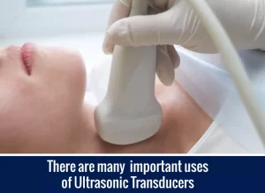
Perhaps the most well-known application of ultrasound is monitoring a pregnancy. Ultrasound provides the means for creating an image of a fetus as it develops inside the mother’s womb. While this is an important use, there are many other applications of ultrasonic transducers in medicine. From diagnostic testing and surgical devices to treating cancer, ultrasonic transducers play a key role in today’s healthcare.
The importance of ultrasonic transducers in healthcare can’t be overstated. Read on to learn more about ultrasonic transducers, including how they were created, how they’re used and the math behind them.
Ultrasound describes sound waves that are above the normal range for human hearing. That means ultrasound comprises very high-frequency sound waves.
This is similar to light. Just as ultraviolet (UV) light is above the frequency for the human eye to see, ultrasound is sound — air pressure waves — that has a frequency too high for the human ear to hear.
Ultrasound is defined typically as anything at 20 kHz — which equates to 20,000 cycles per second — or higher. Medical applications of ultrasound are often at the scale of 10 MHz — 10,000,000 cycles per second — or even higher.
To learn more about ultrasound, take a hearing test. The test starts by presenting an 8 kHz tone, and you can try listening to tones up to 22 kHz. Most people over 25 years old will have trouble hearing anything above 15 kHz. Once you reach the limit on what you can hear, anything higher will be “ultrasound” for you. Keep in mind that in medical applications, the functional frequencies for ultrasound are 100 to 500 times higher than the limit of your own hearing.

To obtain ultrasound on the scale of millions of cycles per second, most medical applications of ultrasound use lead zirconate titanate ceramics, also known as PZT. In case you’re wondering, the P comes from the chemical abbreviation for lead Pb. PZT ceramic is the active part of the piezoelectric ultrasonic transducer used in medical ultrasound. Certain naturally occurring and manmade crystals can also serve as the active part of piezoelectric transducers, but PZT is much more common because it best balances piezoelectric properties and affordability.
A piezoelectric transducer converts electrical energy into sound. It does this because piezoelectric materials change size when a voltage is applied. That means when you apply an alternating current (AC) to piezoelectric material, it starts to vibrate at very high frequencies, which in turn produces high-frequency air pressure waves, or sound waves. Piezoelectric materials can also work in reverse, being used to detect ultrasonic waves and converting the energy from those waves into electricity.
Manufacturing ultrasonic transducers requires sophistication and an understanding of how these instruments will be used in practice. All ultrasonic transducers comprise three components:
1.An active element
2.A backing
3.A matching layer
The active element is the piezoelectric ceramic or crystal that will convert electrical energy into ultrasound energy and back again. The backing is dense and absorbs the energy from the transducer. The matching layer, sometimes known as a wearplate, protects the device from the elements.
Ultrasonic imaging is probably the first thing people think of when they hear the word ultrasound. The imaging is created by using both the echo time of the ultrasound and the Doppler shift of the reflected sound to determine the distance to the targeted internal organ and its movement.
For example, ultrasound is used to image the carotid artery, which is close to the surface of the body. Typically, physicians will be looking for plaque build-up in the artery and the speed of blood flow within the artery.

Ultrasound provides a high-resolution image of the artery at a resolution of about 0.2 millimeters (mm). If you’re working with ultrasound, knowing a little math can help reveal how ultrasound can deliver this fantastic resolution.
The resolution that an ultrasound device provides is a function of a basic equation that governs the wavelength of the resolution. That equation is:
v = fl
This equation translated into words is the velocity of sound in tissue is equal to the frequency of the sound wave multiplied by the wavelength. The Greek letter “l” is the wavelength, “v” is the velocity of sound in tissue — which is about 1,540 meters per second — and “f” is the frequency of the sound wave.
Since the wavelength l determines the resolution, we can rewrite this in terms of l:
l = v/f
When an ultrasound device emits a 7 MHz wave, the resulting resolution is:
l = (1540 meters/second)/(7,000,000 Hz) = 0.22 mm
The higher the frequency, the lower (better) the resolution wavelength; however, the higher the frequency, the more the signal is attenuated. There is an engineering trade-off in designing the right frequency for a particular application.
Let’s return back to our example with the carotid artery. In addition to creating an image of the artery, physicians would like to observe the rate of blood flow, also known as blood flow velocity, within the artery. Ultrasound provides the means for doing this as well.
The ultrasound waves reflect off the target image, which the ultrasound device senses. The reflected waves, however, have a frequency shift away from the frequency emitted originally. This is known as the Doppler Effect.
The Doppler Effect describes the following situations:
– Waves coming toward you from a source that is moving toward you will have a shorter wavelength and thus a higher frequency.
– Waves coming toward to you from a source that’s moving away from you will have a longer wavelength and a corresponding lower frequency.
In medical ultrasound, the Doppler Effect gives rise to a difference between the incident and reflective waves. That difference forms the basis of some additional calculations that determine what’s called the beat frequency. Importantly, the beat frequency is proportional to the blood flow velocity in the artery.
A medical technician can image various locations in the artery and note the blood flow rates. Through these images, the physician has the velocity profile in the artery over time.
Imaging is the most common use of ultrasound in medicine. There are four different models of ultrasound imaging:
1. A-mode: A single transducer scans a single line through the body. The echoes are plotted as a function of depth.
2. B-mode: A linear array of transducers produces a two-dimensional image.
3. M-mode: M refers to motion, in which a quick sequence of B-mode scans provides information on the motion of targeted organs.
4. Doppler mode: This uses the Doppler Effect to measure and visualize blood flow.
The typical applications involve imaging specific areas of the body, including:
Each of these application areas has specific technical demands to provide the best results.
Internal Organs
For example, ultrasound sensors are often used to image internal organs found in the abdomen, such as the liver, kidneys, pancreas and gall bladder. The ultrasound sensors typically use a linear array to create ultrasound frequencies between 2.5 MHz to 7.5 MHz. Based on the calculations made earlier, this provides a resolution of between 0.2 mm and 0.6 mm.
Breasts
Ultrasonic sensors are also used in breast imaging to detect masses or determine a change in their shape. Physicians also use ultrasound to image the thyroid gland to check for masses. These sensors emit a higher frequency sound wave, typically in the range of 7 MHz to 12Mhz.
Cardiovascular System
Cardiologists rely on ultrasound to determine the size and shape of the heart, and to watch the movement of valves inside the heart. Doppler effects can show the direction of blood flow, indicating aspects of regurgitation and leaking. Cardiology sensors operate between 2 MHz to 7.5 MHz. Curiously, vascular sensors looking at blood flow in the body generally rely on a higher frequency range, similar to those used in examining the breast or thyroid.
Eyes
In terms of internal imaging, the application with the highest frequency waveform arises when examining the human eye, particularly prior to cataract surgery. Determining the eye axis length requires as much precision as possible. Correspondingly, the center frequency range for these ultrasound sensors is between 10 MHz and 22MHz.
Uterus
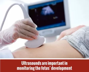
Uterine imagining during pregnancy affords an opportunity to view a developing fetus and track its growth. It can effectively determine the date of conception to within five days. By the time a fetus has developed 18 to 20 weeks, a standard series of images can monitor healthy development of many aspects of the fetus and its environment, including:
In addition to standard two-dimensional image formation, it’s possible to reorganize the images taken and reconstruct a three-dimensional image of the fetus.
For many years, the standard practice for removing kidney stones was via surgical incision. While effective, this comes with all the risks of surgery and general anesthesia. Thanks to today’s technology, doctors use ultrasonic energy to break up kidney stones. This method is not nearly as invasive and does not carry the risks associated with surgery.
To break up kidney stones using ultrasonic energy, A surgeon will make a small incision in the back to insert a nephroscope — a small tube with a light — and find the kidney stone. A metal probe is then inserted through the scope and is guided to make contact with the stone. The metal probe then delivers ultrasonic energy to the stone and breaks it into small pieces. Those pieces are then sucked out by a vacuum.
Some kidney stones respond to what’s called extracorporeal shock wave lithotripsy (ESWL), in which ultrasound waves are focused to the stone from outside the body. Since ESWL doesn’t require an incision, it’s a preferred treatment option. When this option doesn’t work or isn’t indicated, the use of the nephroscope can be the next choice of treatment.
One of the newer uses in ultrasound is as an alternative treatment for prostate cancer. When caught at an early stage, a high-intensity focused ultrasound (HIFU) treatment can be effective.
For the procedure, the surgeon uses a probe to reach the prostate gland via a puncture in the bowel. The probe then releases an HIFU beam that heats and kills the cancer cells. The probe is surrounded by a cooler balloon, which protects the normal cells around it.
Other applications of HIFU include treatment of breast and kidney cancers. In addition, newer techniques use HIFU to heat tissues that are then treated with anti-cancer drugs. Doctors deliver the drugs in liposomes that are sensitive to heat. That way, the doctor can deliver the drug directly to the cancer via the intervention of the HIFU.
Another medical application of ultrasound technology comes in the form of a harmonic scalpel. This scalpel cuts and cauterizes simultaneously, but accomplishes the cauterization by ultrasound rather than via electrical current.
The blade of a harmonic scalpel vibrates 55,500 times per second. Different types of blades — such as curved, hooked, or a combination of curved and hooked — enable the surgeon to perform alternative surgical techniques.
Using a harmonic scalpel, surgeons can perform various surgical procedures. For example, this scalpel is effective in thyroid removal (thyroidectomy). Compared to conventional surgery, the result is a shorter incision length, a reduced operative time and a shorter hospital stay.
For over 25 years, American Piezo (APC International) has been designing and creating the PZT-based piezoelectric transducers the healthcare industry relies on to improve patient outcomes. For piezoelectric ceramic elements designed to fit your specific needs, contact APC.
We create PZT-based piezoelectric transducers in custom shapes — whether they’re based on discs, rings, plates or cylinders — and with exceedingly good quality control. We also can produce other custom piezoelectric ceramics.
At American Piezo, our processing services include designing, pressing, firing, placing custom electrodes, poling, and testing piezoelectric ceramics and devices. We appreciate that custom work requires effective communication with our clients, and we value those working relationships. We know that providing you with first-class service, competitive prices and rapid delivery is what you expect and deserve.
Top Uses of Piezoelectricity in Everyday Applications
Piezo Buzzers vs. Magnetic Buzzers
Principles of Piezoelectric Energy Harvesting
Self Charging Power Cells and Batteries
Overview of Piezoelectric Materials in Energy Harvesting
For more information, call us at 570-726-6961, email us at [email protected] or complete our online contact form. We look forward to hearing from you.